Matching Items (1,503)
Filtering by
- Creators: Marine Biological Laboratory Archives

Description
Specific dendritic morphologies are a hallmark of neuronal identity, circuit assembly, and behaviorally relevant function. Despite the importance of dendrites in brain health and disease, the functional consequences of dendritic shape remain largely unknown. This dissertation addresses two fundamental and interrelated aspects of dendrite neurobiology. First, by utilizing the genetic power of Drosophila melanogaster, these studies assess the developmental mechanisms underlying single neuron morphology, and subsequently investigate the functional and behavioral consequences resulting from developmental irregularity. Significant insights into the molecular mechanisms that contribute to dendrite development come from studies of Down syndrome cell adhesion molecule (Dscam). While these findings have been garnered primarily from sensory neurons whose arbors innervate a two-dimensional plane, it is likely that the principles apply in three-dimensional central neurons that provide the structural substrate for synaptic input and neural circuit formation. As such, this dissertation supports the hypothesis that neuron type impacts the realization of Dscam function. In fact, in Drosophila motoneurons, Dscam serves a previously unknown cell-autonomous function in dendrite growth. Dscam manipulations produced a range of dendritic phenotypes with alteration in branch number and length. Subsequent experiments exploited the dendritic alterations produced by Dscam manipulations in order to correlate dendritic structure with the suggested function of these neurons. These data indicate that basic motoneuron function and behavior are maintained even in the absence of all adult dendrites within the same neuron. By contrast, dendrites are required for adjusting motoneuron responses to specific challenging behavioral requirements. Here, I establish a direct link between dendritic structure and neuronal function at the level of the single cell, thus defining the structural substrates necessary for conferring various aspects of functional motor output. Taken together, information gathered from these studies can inform the quest in deciphering how complex cell morphologies and networks form and are precisely linked to their function.
ContributorsHutchinson, Katie Marie (Author) / Duch, Carsten (Thesis advisor) / Neisewander, Janet (Thesis advisor) / Newfeld, Stuart (Committee member) / Smith, Brian (Committee member) / Orchinik, Miles (Committee member) / Arizona State University (Publisher)
Created2013

Description
In somatic cells, the mitotic spindle apparatus is centrosomal and several isoforms of Protein Kinase C (PKC) have been associated with the mitotic spindle, but their role in stabilizing the mitotic spindle is unclear. Other protein kinases such as, Glycogen Synthase Kinase 3â (GSK3â) also have been shown to be associated with the mitotic spindle. In the study in chapter 2, we show the enrichment of active (phosphorylated) PKCæ at the centrosomal region of the spindle apparatus in metaphase stage of 3T3 cells. In order to understand whether the two kinases, PKC and GSK3â are associated with the mitotic spindle, first, the co-localization and close molecular proximity of PKC isoforms with GSK3â was studied in metaphase cells. Second, the involvement of inactive GSK3â in maintaining an intact mitotic spindle was shown. Third, this study showed that addition of a phospho-PKCæ specific inhibitor to cells can disrupt the mitotic spindle microtubules. The mitotic spindle at metaphase in mouse fibroblasts appears to be maintained by PKCæ acting through GSK3â. The MAPK pathway has been implicated in various functions related to cell cycle regulation. MAPKK (MEK) is part of this pathway and the extracellular regulated kinase (ERK) is its known downstream target. GSK3â and PKCæ also have been implicated in cell cycle regulation. In the study in chapter 3, we tested the effects of inhibiting MEK on the activities of ERK, GSK3â, PKCæ, and á-tubulin. Results from this study indicate that inhibition of MEK did not inhibit GSK3â and PKCæ enrichment at the centrosomes. However, the mitotic spindle showed a reduction in the pixel intensity of microtubules and also a reduction in the number of cells in each of the M-phase stages. A peptide activation inhibitor of ERK was also used. Our results indicated a decrease in mitotic spindle microtubules and an absence of cells in most of the M-phase stages. GSK3â and PKCæ enrichment were however not inhibited at the centrosomes. Taken together, the kinases GSK3â and PKCæ may not function as a part of the MAPK pathway to regulate the mitotic spindle.
ContributorsChakravadhanula, Madhavi (Author) / Capco, David G. (Thesis advisor) / Chandler, Douglas (Committee member) / Clark-Curtiss, Josephine (Committee member) / Newfeld, Stuart (Committee member) / Arizona State University (Publisher)
Created2012

Description
Background
Drosophila melanogaster has been established as a model organism for investigating the developmental gene interactions. The spatio-temporal gene expression patterns of Drosophila melanogaster can be visualized by in situ hybridization and documented as digital images. Automated and efficient tools for analyzing these expression images will provide biological insights into the gene functions, interactions, and networks. To facilitate pattern recognition and comparison, many web-based resources have been created to conduct comparative analysis based on the body part keywords and the associated images. With the fast accumulation of images from high-throughput techniques, manual inspection of images will impose a serious impediment on the pace of biological discovery. It is thus imperative to design an automated system for efficient image annotation and comparison.
Results
We present a computational framework to perform anatomical keywords annotation for Drosophila gene expression images. The spatial sparse coding approach is used to represent local patches of images in comparison with the well-known bag-of-words (BoW) method. Three pooling functions including max pooling, average pooling and Sqrt (square root of mean squared statistics) pooling are employed to transform the sparse codes to image features. Based on the constructed features, we develop both an image-level scheme and a group-level scheme to tackle the key challenges in annotating Drosophila gene expression pattern images automatically. To deal with the imbalanced data distribution inherent in image annotation tasks, the undersampling method is applied together with majority vote. Results on Drosophila embryonic expression pattern images verify the efficacy of our approach.
Conclusion
In our experiment, the three pooling functions perform comparably well in feature dimension reduction. The undersampling with majority vote is shown to be effective in tackling the problem of imbalanced data. Moreover, combining sparse coding and image-level scheme leads to consistent performance improvement in keywords annotation.
ContributorsSun, Qian (Author) / Muckatira, Sherin (Author) / Yuan, Lei (Author) / Ji, Shuiwang (Author) / Newfeld, Stuart (Author) / Kumar, Sudhir (Author) / Ye, Jieping (Author) / Biodesign Institute (Contributor) / Center for Evolution and Medicine (Contributor) / College of Liberal Arts and Sciences (Contributor) / School of Life Sciences (Contributor) / Ira A. Fulton Schools of Engineering (Contributor)
Created2013-12-03

Description
Background
Multicellular organisms consist of cells of many different types that are established during development. Each type of cell is characterized by the unique combination of expressed gene products as a result of spatiotemporal gene regulation. Currently, a fundamental challenge in regulatory biology is to elucidate the gene expression controls that generate the complex body plans during development. Recent advances in high-throughput biotechnologies have generated spatiotemporal expression patterns for thousands of genes in the model organism fruit fly Drosophila melanogaster. Existing qualitative methods enhanced by a quantitative analysis based on computational tools we present in this paper would provide promising ways for addressing key scientific questions.
Results
We develop a set of computational methods and open source tools for identifying co-expressed embryonic domains and the associated genes simultaneously. To map the expression patterns of many genes into the same coordinate space and account for the embryonic shape variations, we develop a mesh generation method to deform a meshed generic ellipse to each individual embryo. We then develop a co-clustering formulation to cluster the genes and the mesh elements, thereby identifying co-expressed embryonic domains and the associated genes simultaneously. Experimental results indicate that the gene and mesh co-clusters can be correlated to key developmental events during the stages of embryogenesis we study. The open source software tool has been made available at http://compbio.cs.odu.edu/fly/.
Conclusions
Our mesh generation and machine learning methods and tools improve upon the flexibility, ease-of-use and accuracy of existing methods.
ContributorsZhang, Wenlu (Author) / Feng, Daming (Author) / Li, Rongjian (Author) / Chernikov, Andrey (Author) / Chrisochoides, Nikos (Author) / Osgood, Christopher (Author) / Konikoff, Charlotte (Author) / Newfeld, Stuart (Author) / Kumar, Sudhir (Author) / Ji, Shuiwang (Author) / Biodesign Institute (Contributor) / Center for Evolution and Medicine (Contributor) / College of Liberal Arts and Sciences (Contributor) / School of Life Sciences (Contributor)
Created2013-12-28
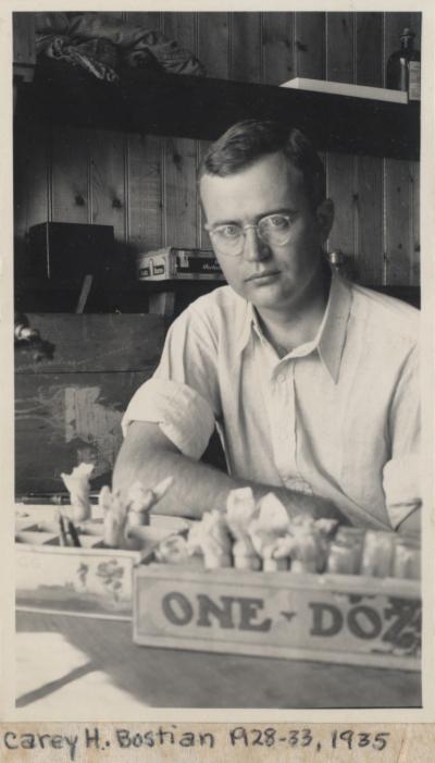
ContributorsMarine Biological Laboratory Archives (Publisher) / Arizona Board of Regents (Publisher)
Created1935
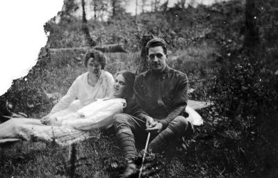
ContributorsHuettner, Alfred F. (Alfred Francis), b. 1884 (Creator) / Marine Biological Laboratory Archives (Publisher)
Created1918
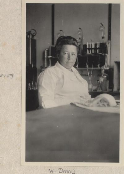
ContributorsMarine Biological Laboratory Archives (Publisher)
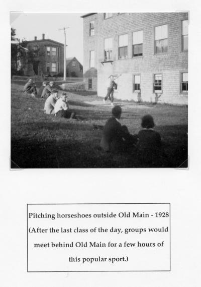
ContributorsHuettner, Alfred F. (Alfred Francis), b. 1884 (Creator) / Marine Biological Laboratory Archives (Publisher)
Created1928
ContributorsHuettner, Alfred F. (Alfred Francis), b. 1884 (Creator) / Marine Biological Laboratory Archives (Publisher)
Created1918
ContributorsHuettner, Alfred F. (Alfred Francis), b. 1884 (Creator) / Marine Biological Laboratory Archives (Publisher)